

M Ozge Tepe
Istanbul University, Turkey
Title: Histopathological findings in surgically resected treatment-resistant epilepsy cases
Biography
Biography: M Ozge Tepe
Abstract
Statement of the Problem: In treatment-resistant epilepsy, different etiologies, histomorphological and immunohistochemical features, and diseases are included. Hippocampal sclerosis and focal cortical dysplasia are the most common histopathological diagnosis while tumor, vascular malformation, encephalitis, and glial scar are featured in decreasing frequency. Hippocampal sclerosis and focal cortical dysplasia are histopathologically classified according to the International League Against Epilepsy (ILAE) classifications. Molecular genetic studies in recent years have been effective in determining targeted therapies in patients who do not respond to antiepileptic drugs. mTOR pathway and immune system activation have been shown to play a role in epileptogenesis. To determine the incidence of different etiologies in the treatment-resistant epilepsy patients and find out histomorphological and immunohistochemical features and to demonstrate the relation between the ILAE subtypes and the clinical features and try to predict the prognosis of the patients were main purposes in a neuropathological examination of our surgically resected treatment-resistant epilepsy cases.
Methodology & Theoretical Orientation: In addition to immunohistochemistry (NeuN, Neurofilament-H, CD34, GFAP, IDH-1, and Olig-2) was performed in the diagnostic process, pS6 was used to demonstrate mTOR pathway activation in FCD cases and CD3, CD8, Iba-1 antibodies were applied to demonstrate neuroinflammation in HS cases.
Findings: Statistical analysis of HS and FCD, were the most frequent histological findings, revealed a significant difference in age of seizure onset, epileptic seizure duration, surgical age, gender status, and Engel classification. pS6 expression was observed in dysmorphic neurons and balloon cells in the cases of FCD type II while lymphocyte infiltration was seen in all HS cases.
Conclusion & Significance: Significant pS6 expression in FCD type II indicates that mTOR pathway inhibitors may be involved in the treatment of epilepsy. In HS cases, no statistical significant pathological feature to predict efficacy of immunomodulating therapy in a special subgroup has been identified.
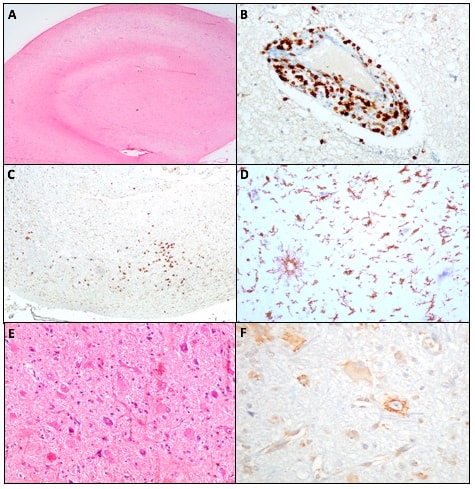
Figure: (A) Hippocampal sclerosis showing neuronal loss in CA1 and CA4. (B) Perivascular CD3 immunopositivity. (C) Paranchymal CD3 immunopositivity. (D) Microglial cells showing Iba-1 immunopositivity. (E) Focal cortical dysplasia containing dysmorphic and balloon cells. (F) pS6 immunopositivity in dysmorphic cells and baloon cells.