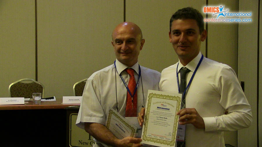
Tuncer Kutlu
Ankara University, Turkey
Title: Pathomorphological and ımmunohistochemical evaluation of osteoid osteoma lıke osteoblastoma in proximal humerus of a dog: an unusal case
Biography
Biography: Tuncer Kutlu
Abstract
Osteoid osteoma and osteoblastoma are uncomon benign bone tumor which originated from osteoblasts in animals. There are two clinic reports including mandible in a cat and in humerus in a dog. Both tumors have also similar characteristics. Macroscopically, osteoid osteoma is smaller and extend to soft tissues. Histopathologically, osteoid osteoma is composed of cellulary uniformity, sclerotic bone and vascular osteoid tissue. However, osteoblastoma has more extensive lysis and sclerotic areas. In the case, both tumor characteristics were present. By using some markers, differentiation of the tumors was aimed. 12 years old mix breed female dog was submitted into clinic with request of progressive loss of motion and swelling in proximal forelimb. During therapy, animal was died and cadaver was sent to Department of Pathology. After macroscopical examination, samples were taken and fixed in %10 formalin. For histopathology, hematoksilen-eozin, Masson’s trichrome and Alizerin Red S staining methods were used. For differentiation, ABC-P was applicated to adhesive section by using BMP6, S100, vimentin ve p53 markers. Macroscopically, proximal humerus was swollen being 12 cm in diameter. Cut section was generally lytic within bone residues. Soft tissue was haemorrhagic and edematouse. Microscopically, osteoblasts and hypervascular nidus were surrounded large sclerotic and mineralized bone. Large lytic areas were attended. Masson’s trichrome differentiated sclerosis and Alizerin Red S showing osteoid matrix. Immunohistochemically, BMP6 was moderetely reacted in osteoid matrix and S100 was modeately reacted with osteoblasts in nidus. Vimentin gave weakly positive reaction in fibrocytes. P53 was negative in osteoblasts. In conclusion,tumor resembling to osteoblastoma in terms of distribution is a rare case in animals. Immunohistochemistry is not evaluated beforeones. The case show that BMP6 and S100 are usefull in presence of osteoid matrix and differentiation. P53 proven its to be malignant potential.

